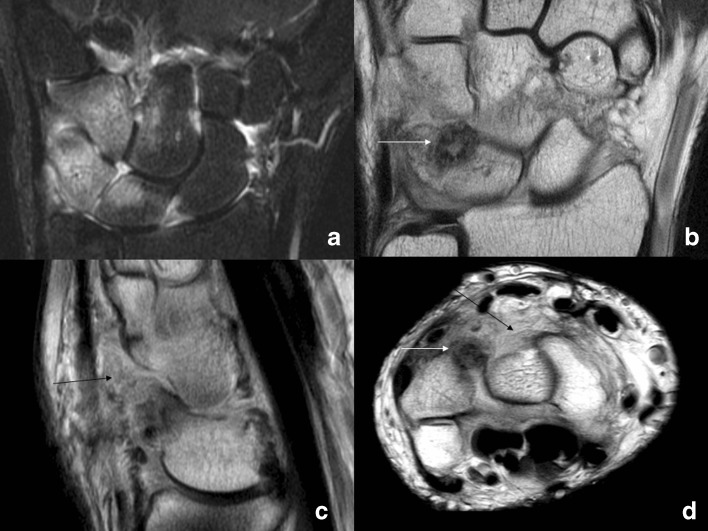
Fig. 2.
MRI of the left wrist performed in August of 2010. a On the coronal STIR, there is bone marrow edema in the lunate, triquetrum, hamate, and capitate, as well as synovitis in the radiocarpal, midcarpal, and carpometacarpal joints. The coronal (b), sagittal (c), and axial (d) proton density images show a round area of decreased signal (white arrows) in the dorsal aspect of the triquetrum corresponding to the “cyst” that had been curetted and grafted previously. There is synovial thickening (black arrows) in the soft tissues dorsally.

