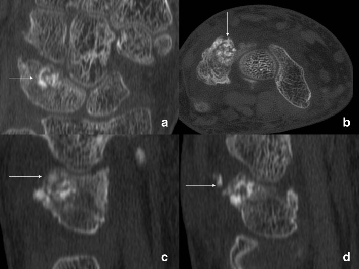
Fig. 3.
CT of the left wrist performed in September of 2010. The coronal (a), axial (b), and sagittal images (c, d) show a round lesion in the dorsal aspect of the triquetrum packed with incorporated bone graft (arrows) with some graft in the soft tissues dorsally. Fusion of the triquetrum and lunate is seen on the coronal view (a).

