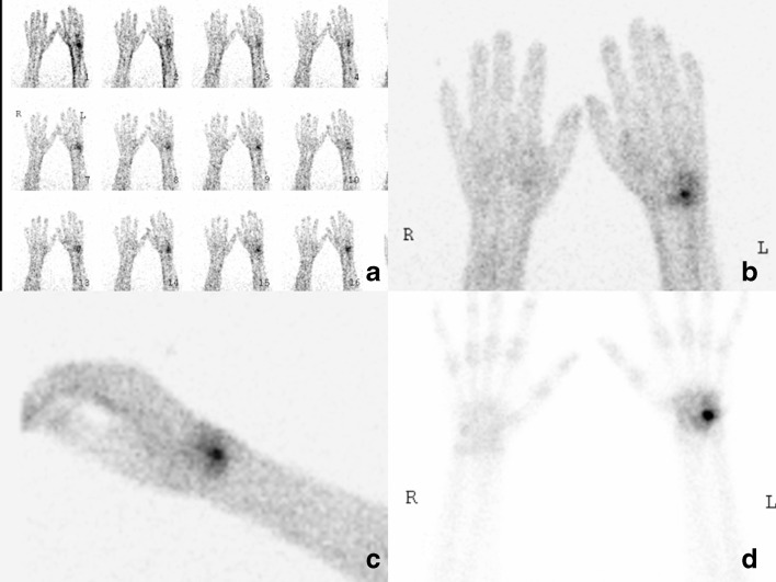
Fig. 4.
Three-phase bone scan of the wrists and hands performed in 2010. The intravenous injection of technetium 99m MPD was done in the right elbow. On the dynamic flow study (a), there is increased flow to the left wrist. The blood pool scans (b, c) show focal increased blood pool in the ulnar and dorsal aspects of the wrist around the left triquetrum. The delayed static image (d) shows a round focal area of high uptake in the left triquetrum typical of an osteoid osteoma. There is mild surrounding increased uptake consistent with synovitis. There is no diffuse periarticular uptake suggestive of CRPS.

