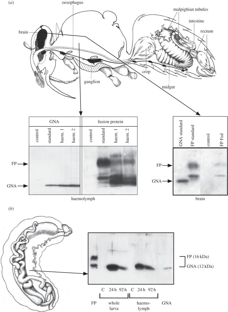
Figure 3.
Immuno-assay by western blotting demonstrates internalization of Hv1a/GNA in adult honeybee tissues. Bands of GNA (12 kDa) and Hv1a/GNA (FP; 16 kDa) are indicated. (a) Diagram of adult honeybee showing the presence of GNA and fusion protein Hv1a/GNA (FP) in both the haemolymph and brain after feeding solutions containing proteins. Insects were fed 100 µg GNA or Hv1a/GNA, and haemolymph or brain tissue was collected after 24 h for analysis. (b) Diagram of larval honeybee showing that Hv1a/GNA (FP) is degraded after ingestion; larvae were dosed with 100 µg Hv1a/GNA per larva and haemolymph was collected after 24 h for analysis.

