Figure 7.

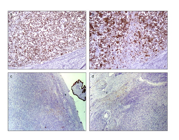
Immunohistochemical staining of left ovary. a: p63 positivity (100 x); b: High molecular weight cytokeratin CK5/6 positivity (100 x); c: CK7 negativity (40 x); d: CK20 negativity (40 x)

Immunohistochemical staining of left ovary. a: p63 positivity (100 x); b: High molecular weight cytokeratin CK5/6 positivity (100 x); c: CK7 negativity (40 x); d: CK20 negativity (40 x)