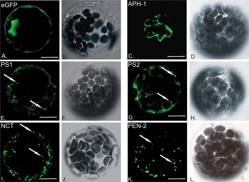
Fig. 2.
Cellular localization of γ-secretase subunits. Genetic constructs coding for γ-secretase subunits fused with eGFP were introduced to Arabidopsis thaliana leaf mesophyll protoplasts for transient expression and analysed with confocal microscope. (A) eGFP fluorescence alone was used as control; (C) AtAPH-1–GFP signal is visible in reticular structures; (E) AtPS1–GFP and (G) AtPS2–GFP signal marks vesicular compartments and reticular structures; (I) AtNCT–GFP and (K) AtPEN-2–GFP fluorescence visible mostly in vesicles. Transmitted light images of respective cells are presented next to fluorescent sections (B, D, F, H, J, L). White arrows point to exemplary vesicles. Bar, 10 µm.

