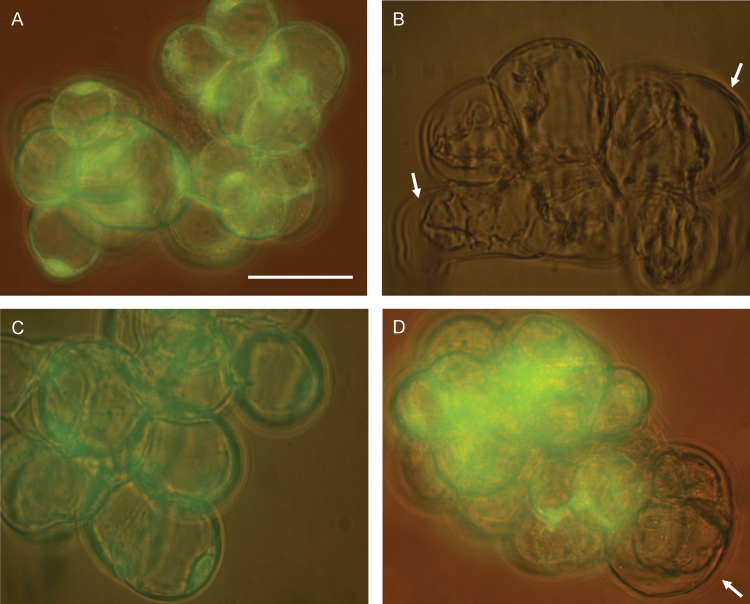
Fig. 4.
Representative field images showing changes in the morphology of light-grown Arabidopsis cells under control conditions (A), after the 30min treatment with 0.5 μM RB (B) and 500 μM H2O2 (C), and after the 90min treatment at 1800 μE m−2 s−1 (D). Bright green fluorescence of fluorescein diacetate following excitation with UV radiation in A, C, and D indicates that Arabidopsis cells remain viable after the stress treatment. Arrows in B and D illustrate some of the gaps between the cell wall and the plasma membrane after the induction of PCD. Scale bars=50 μm.

