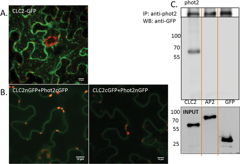
Fig. 8.
Phot2 association with light chain2 of clathrin. (A) Light chain2 of clathrin (AtCLC2) transiently expressed in N. benthamiana. (B) BiFC technique used to examine the association between phot2 and CLC2. (C) Co-immunoprecipitation assay: the microsomal fraction proteins isolated from N. benthamiana leaves were subjected to immunoprecipitation (using Atphot2 antibody) and then to western blot. The upper blot shows phot2 (after use of an anti-Atphot2 antibody) in Nicotiana leaves transiently expressing phot2I plus CLC2–GFP, GFP–AP2μ, and GFP respectively. The lower blot shows a single band corresponding to CLC2–GFP after anti-GFP staining. As control (input), microsomal fractions (100 μg) were also immunoblotted before immunoprecipitation; the bands were identified using anti-GFP antibody. Numbers represent molecular weights (kDa) of the protein marker (Thermo Scientific).

