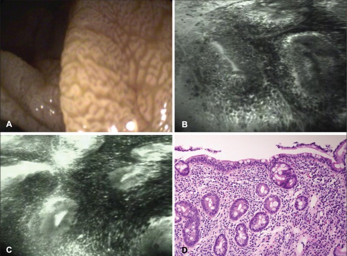
Figure 1).
A White-light endoscopy revealing mosaic and nodular pattern. B and C Total villous atrophy with collars of enterocytes around the crypt openings and an increased number of intraepithelial lymphocytes and leakage of fluorescein. D Corresponding histological specimen reveals a Marsh IIIb focally IIIc lesion with total absence of villi and numerous intraepithelial lymphocytes (hematoxylin and eosin stain, original magnification ×20)

