Figure 2).

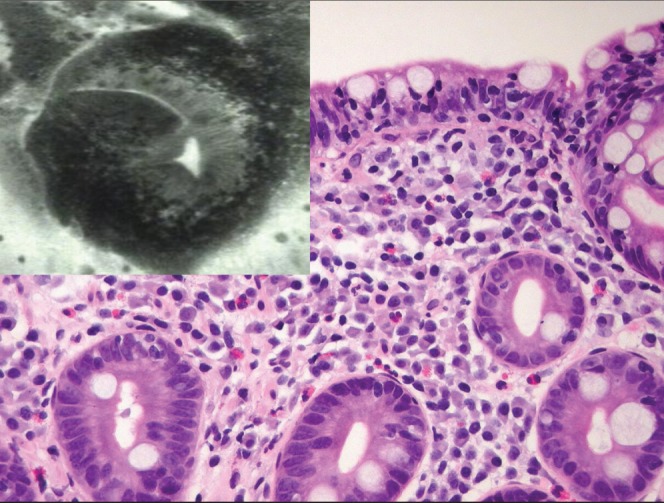
Villous atrophy and the base of the crypts appears as roundish glands resembling colonic daisy crypts. Corresponding histological specimen reveals a Marsh IIIb focally IIIc lesion. Hematoxylin and eosin stain, original magnification ×20

Villous atrophy and the base of the crypts appears as roundish glands resembling colonic daisy crypts. Corresponding histological specimen reveals a Marsh IIIb focally IIIc lesion. Hematoxylin and eosin stain, original magnification ×20