Figure 3).

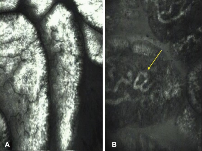
Confocal endomicroscopy images of normal duodenal mucosa. A Villi appear regular, ‘fingerlike’ with goblet cells between the enterocytes. B Endomicroscopy especially highlights capillaries within the lamina propria (arrow)

Confocal endomicroscopy images of normal duodenal mucosa. A Villi appear regular, ‘fingerlike’ with goblet cells between the enterocytes. B Endomicroscopy especially highlights capillaries within the lamina propria (arrow)