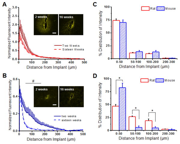
Figure 4.

Blood-brain barrier permeability (IgG) around implanted microelectrodes in the rat and the the mouse model. (A) In the rat model, similar IgG+ immunoreactivity was observed at two weeks (solid red) and sixteen weeks (dashed red). (B) In contrast, significantly more IgG+ immunoreactivity was noted from 50 to 200 μm away from the implanted device at two weeks post implantation in the mouse model. (C) Similar IgG+ distribution profiles were observed in both the rat and the mouse model at two weeks. (D) By sixteen weeks, however, a more diffuse/widespread IgG+ distribution profile was noted in the rat model (0-200 μm) compared to the mouse model. *p<0.05; Scale = 100 μm; n = 4-7 animals for each cohort
