
Figure 4.
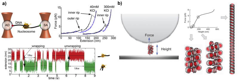
Micro and nano methods for understanding chromatin dynamics and chromosome organization. a) Optical tweezers experiment on a single nucleosome and force extension curves of the nucleosome in different salt concentrations106. The lower panel shows the nucleosome flipping between a wrapped and unwrapped state. b) Magnetic tweezers setup (left panel), where a single heterochromatin fibre can be pulled apart into different conformations (right panel)107. The different points on the force-extension curve show as the pulling force is increased, the fibre begins to unravel in a manner similar to a Hookian spring (solenoid shape), which keeps the DNA both condensed and accessible.