Fig. 2.

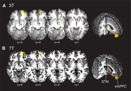
MR Images showing the connection (red) between vmPFC (yellow) and STN (green) for 3 Tesla (A) and 7 Tesla (B) images. Background images are anatomical scans acquired at a 3T Philips (T1) and 7T Siemens (MP2RAGE) scanner

MR Images showing the connection (red) between vmPFC (yellow) and STN (green) for 3 Tesla (A) and 7 Tesla (B) images. Background images are anatomical scans acquired at a 3T Philips (T1) and 7T Siemens (MP2RAGE) scanner