Fig. 2.

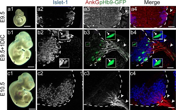
Whole embryos cultured for one day upon electroporation exhibit a normal development. a1, b1, c1 E9.5 embryos which were electroporated and then maintained for one day in culture (E9.5 + 1DIC, b1) appeared to develop normally when compared to control embryos observed at E10.5 (c1) or E9.5 (a1): they displayed an increase in their size, the development of forelimb buds (single asterisk) and the emergence of hindlimb buds (double asterisks). a2, b2, c2 Immunolabeling of Islet1 on coronal sections of the spinal cord in (a1, b1, c1) also reveals the increased number of Islet1+ MNs in the spinal cord ventral horn after one day in culture (compare b2, c2 and a2; dashed lines delimit the spinal cord). a3, b3, c3 Immunolocalization of AnkG (with rabbit anti-AnkG) confirms the extent of its distribution along developing motor axons (arrowheads) in electroporated GFP+ MNs as in E10.5 control embryos (compare b3, c3 and a3 as well as Fig. 1d2). Insets in b are enlargements of dashed boxed areas, highlighting the MN identity of GFP+ cells (arrows, n = 3; 32 neurons). Scale bars represent 1 mm in a1, b1, c1, 25 μm in a2–a4, b2–b4, c2–c4 and 5 μm in the enlargements in b
