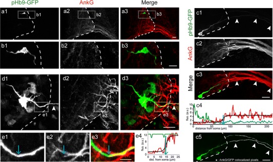
Fig. 3.

AnkG is never expressed in migrating MNs and its expression is first detected in the axon of morphologically differentiated MNs. a, b Bipolar MNs electroporated with the Hb9-GFP plasmid did not express AnkG. No AnkG immunolabeling (with rabbit anti-AnkG) (a2, b2) was detected neither on the trailing nor on the leading process (n = 6; 49 neurons) of immature bipolar GFP+ MNs (a1, b1). b1–b3 Enlargements of the dashed boxes in (a1–a3), respectively. c–e More differentiated MNs expressed AnkG along their axon. More differentiated GFP+ MNs (c1, d1) with an axon (arrowheads) crossing the spinal cord boundary (dashed lines) expressed AnkG (c2, d2). e1–e3 Enlargements of the dashed box in (d1–d3), respectively. e4, c4 Quantification of AnkG (red) and GFP (green) fluorescence along motor axons shown in e1 and c1, respectively. Black curves are theoretical sigmoids fitted to the AnkG fluorescence profile. Blue arrows indicate the starting point of AnkG expression, as being the inflexion point of the sigmoid curve. c5 Distribution of AnkG+ pixels (in white) colocalizing with GFP (in green) in C3, along the GFP+ axon (from c3). Scale bars represent 25 μm in a, b, c and 5 μm in e
