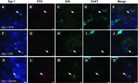
Fig. 5.

Immediate early gene expression in the Parkinsonian rat. a–e Fluorescent images showing an Egr-1-positive PPN neuron (a, arrow) that was retrogradely labeled from the STN (b), but not the GiN (c), and was non-cholinergic (d; merged image in e). f–j Fluorescent images of an Egr-1-positive PPN neuron (f, arrow) that was not retrogradely labeled from the STN (g), but was labeled from the GiN (h; i, also non-cholinergic; merged image in j). K–O Fluorescent images of an Egr-1-positive PPN neuron (k arrow) that projects to both the STN (l) and GiN (m; n, also non-cholinergic; merged image in o). Scale bars 10 μm
