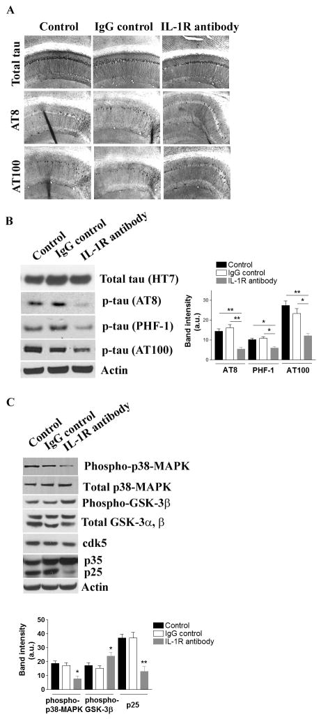
Figure 4. Suppressing IL-1 signaling attenuates tau pathology in the 3xTg-AD mice.
(A) Tau pathology is attenuated by blocking IL-1 signaling. Representative immunohostochemical staining with various tau antibodies; HT7 – total tau, AT8 – phosphorylated tau at Ser 199 and Ser 202, and AT100 – phosphorylated tau at Ser 212 and Thr 214. (B) Immunoblot and densitometric analyses of changes in the steady-state levels of phosphorylated tau in the brain. Each bar is expressed as mean ± S.E.M. (n=10 for control and IgG control, and n=8 for anti-IL-1R treatment), and *p<0.05 or **p<0.01 compared to control and IgG control groups. More immunoblot data for phospho-tau are found in Suppl. Fig. 2D. (C) Suppressing IL-1 signaling results in decreased activations of tau kinases. Immunoblot and densitometric analyses of the steady-state levels of GSK-3β, cdk5/p35/p25 and p38-MAPK. Each bar is expressed as mean ± S.E.M. (n = 10 for control and IgG control, and n=8 for anti-IL-1R treatment), and *p<0.05 or **p<0.01 compared to control and IgG control groups.

