4.
AIM is a new tool to expose image metadata and make it accessible for a variety of applications.
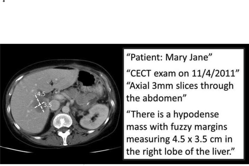
4a. Image metadata. This shows an example image and excerpts from the radiology report. The graphical symbols drawn by the radiologist on the image (“markups” indicating measurements of a lesion—quantitative data) and the statements in the report about the patient, type of exam, technique, date, imaging observations, and anatomic localization (semantic data) collectively comprise the image metadata (“annotation”). These image metadata, if stored in a standardized, machine-accessible format, greatly enable many computer applications to help radiologists in their daily work.
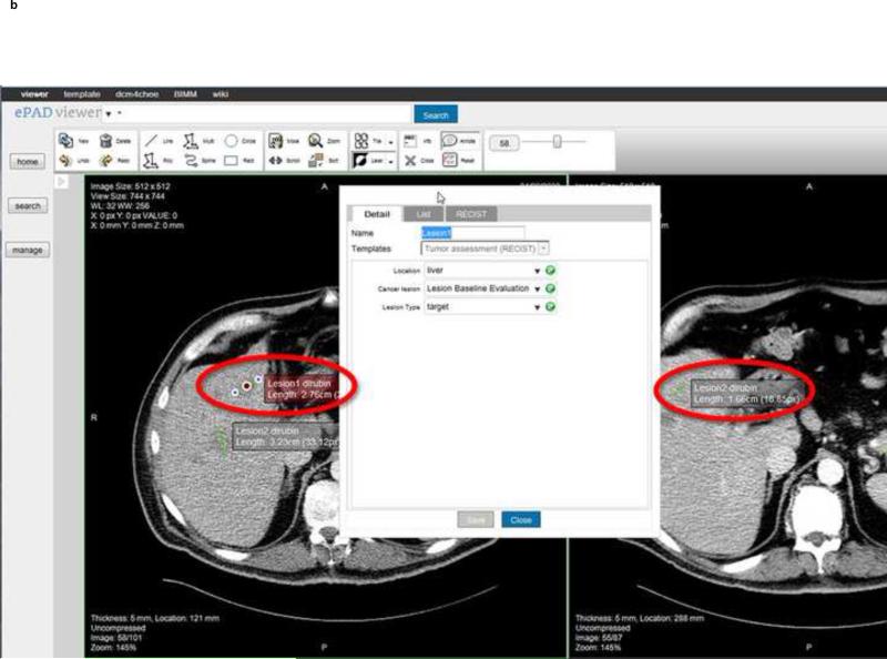
4b. ePAD rich Web client. The ePAD application provides a platform-independent and thin client implementation of an AIM-compliant image viewing workstation. Information about lesions that are marked up and reported by radiologists is captured and stored in AIM XML (or DICOM-SR). User-definable templates capture semantic information about lesions, such as shown in this case for oncology reporting, the type of lesion (target), anatomic location (liver), and type of imaging exam (baseline evaluation).
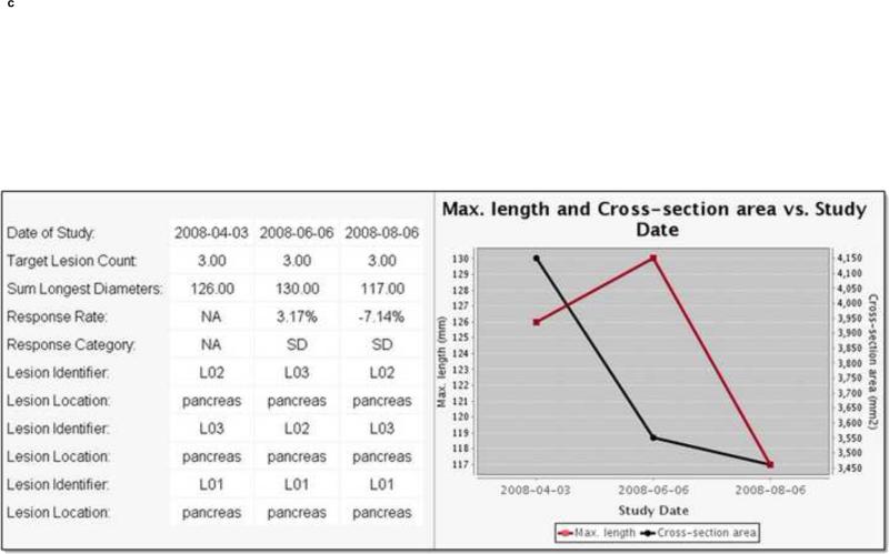
4c. Radiology image information summarization application levering the utility of AIM-encoded image metadata. A cancer lesion tracking application has queried AIM annotations created on different imaging studies (in this case, from 3 studies on 4/3/08, 6/6/08, and 8/6/08). The application automatically calculates the sum of each target lesion measured on each imaging study date and summarizes the results in a table (left) and graph (right). Using metadata from the AIM annotations, the application also displays alternative response measures such as maximum length (red line) or cross-sectional area (black line) of the measured lesions.


