
Figure 5.
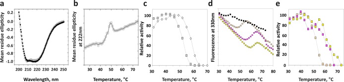
Structural stability of the isolated phosphatase domain of PHLPP. (a) Circular dichroism (CD) spectra for PHLPP1 (■) and PHLPP2 (△) phosphatase domains, isolated from bacteria. (b) The phosphatase domain of PHLPP2 was subjected to increasing temperatures, and variations of the mean residue ellipticity at 222 nm (by CD) were recorded. (c) Activity of the phosphatase domain of PHLPP1 (empty diamonds) or PHLPP2 (gray squares) was measured by fluorescence at different temperatures using DiFMUP as the substrate. (d) Phosphatase domain PHLPP2 treated with EDTA (black squares) and subsequently incubated with MnCl2 (orange squares), MgCl2 (yellow squares), or CaCl2 (pink squares) was subjected to increasing temperatures, and fluorescence emission at 330 nm was recorded. (e) Activity of PHLPP2 incubated with MnCl2 (orange squares), MgCl2 (yellow squares), or CaCl2 (pink squares) was measured by fluorescence at different temperatures using DiFMUP as the substrate.