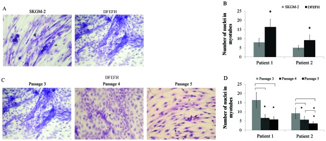
Figure 4.
Fusion potential of cultured myoblasts in different media. After passage 3, cells cultured in 2 different media were seeded in duplicate into 96-well plates, and when 80% confluence was obtained, the culture medium was switched to fusion medium [Dulbecco’s modified Eagle’s medium (DMEM), 2% fetal bovine serum (FBS), 25 μmol/l insulin] and changed every 3 days. After 11 days, cells were fixed and stained with Wright’s eosin methylene blue solution. The number of nuclei in the 6 largest myotubes was counted in each well. Additionally, cells cultured in DFEFH medium were subjected to higher passages and their fusogenic potential was assessed in a similar manner. (A) Comparison of the fusogenic potential of myoblasts cultured in skeletal muscle cell growth medium-2 (SKGM-2) and DFEFH medium at passage 3. Representative results of 2 independent experiments are shown. (B) The average number of nuclei in the largest myotubes created from myoblasts expanded in SKGM-2 and DFEFH medium at passage 3, n=2 (2 different donors); *p<0.05. (C) The fusion potential of myoblasts in prolonged cultured in DFEFH medium. Representative results of 2 independent experiments are shown. (D) The average number of nuclei in the largest myotubes created from myoblasts expanded in DFEFH medium at passages 3, 4 and 5, n=2 (2 different donors); *p<0.05.

