
FIGURE 3.
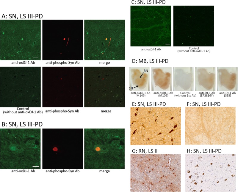
Immunohistochemical distribution of oxDJ-1 in the human midbrain. (A–C) Confocal images of oxDJ-1 in dopaminergic nigral neuronal cells. A section containing the SN was immunostained with a mAb against oxDJ-1 and a polyclonal antibody against phosphorylated α-synuclein and then visualized using fluorescence confocal microscopy. (A) Staining with only the secondary antibody is shown in the lower panel. (B) Immunoreactivity of the anti–oxDJ-1 mAb was widely distributed throughout the cell bodies and neurites in dopaminergic neurons of the SN. The areas labeled with anti–oxDJ-1 and anti-phosphorylated α-synuclein (indicative of LBs) antibodies were colocalized (A). Higher magnification is shown in (B). At the periphery of the SN, oxDJ-1 IR was clearly observed in neuron processes (C). (D) Oxidized DJ-1 IR was present throughout the midbrain; particularly high levels were observed in the SN and red nucleus (RN). No staining with only the secondary antibody and less staining with the anti–DJ-1 mAbs (clones EP2816Y and 3E8) are shown. (E–H) Staining results at higher magnification of the SN (E, F, H) and RN (G) using the oxDJ-1 mAb (E–G) and only the secondary antibody (H). There was IR of cell bodies and processes of neuromelanin-containing neurons with the oxDJ-1 mAb (E); there was no staining with the secondary antibody alone (H). At the periphery of the SN, oxDJ-1 IR was evident in neuron processes (F). In the RN, there was oxDJ-1 IR of glial cells and neuron processes (G). Scale bars = (A, C, E) 50 μm; (B, F) 20 μm. LB I, patient with scattered LBs without cell loss; LS II, patient with LBs and cell loss but without clinical parkinsonism or dementia; LS III–PD, patients with LB and PD; MB, midbrain.