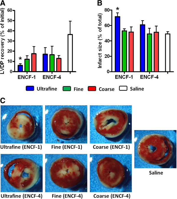
Figure 6.

Post ischemia-reperfusion cardiac end points in mice at 24 h post-exposure to ENCF-1 and ENCF-4 PM (100 μg) by oropharyngeal aspiration. (A) recovery of LVDP, expressed as a percentage of the initial pre-ischemic LVDP, measured at 1 h of reperfusion after 20 min of ischemia. (B) infarct size, expressed as a percentage of the total ventricle area, measured at 2 h of reperfusion after 20 min of ischemia. (C) representative images of mouse heart sections stained with TTC. Red-stained areas indicate viable tissue and negatively stained (white) areas indicate infracted tissue. Data are means ± SEM (n = 4–5 in each group). *p < 0.05 compared with the saline-exposed negative control group.
