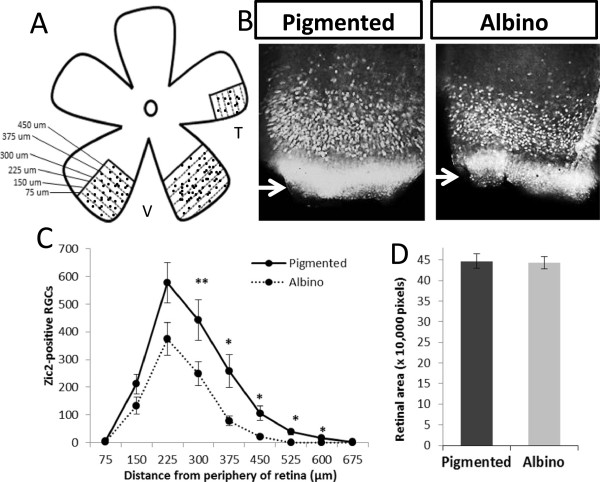
Figure 1.

The density of Zic2+ retinal ganglion cells, but not their distribution, is changed at embryonic day 15.5 in the albino retina. (A) Cartoon depicting the method for counting Zic2+ retinal ganglion cells (RGCs) in 75 μm sectors from the periphery towards the center of the ventrotemporal (VT) retina. V, ventral; T, temporal. Black dots represent Zic2+ RGCs and dotted lines represent delineations of sectors. (B) Fluorescent images of retinal flat mounts immunostained with a Zic2 antibody at embryonic day 15.5. Zic2 is expressed in VT RGCs. (C) The number of Zic2+ cells in each sector of pigmented (n = 6) and albino (n = 6) VT retinae. In each sector, the number of Zic2+ cells is lower in albino compared to pigmented retina. However, the peak and distribution of Zic2+ cells are similar in the pigmented and albino retina. (D) The area of retinal flatmount does not differ in the pigmented (n = 11) and albino (n = 9) eyes at embryonic day 15.5. For area measurements, 1 pixel = 4.2734 μm2. Error bars indicate SEM. Two-tailed unpaired t-test: *P < 0.05, **P < 0.01.
