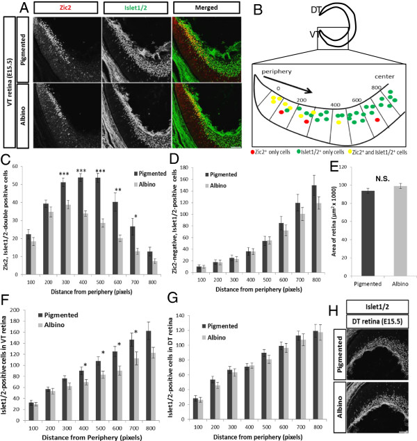
Figure 2.

The expression of Zic2 and Islet1/2 in the pigmented and albino retina at embryonic day 15.5. (A) Frontal sections through ventrotemporal (VT) retina immunostained with antibodies against Islet1/2 (postmitotic retinal ganglion cells (RGCs), green) and Zic2 (ipsilateral RGCs, red) at embryonic day (E)15.5. Zic2+ RGCs are situated in the most peripheral region of VT retina. (B) (top) Cartoon depicting region of retina where Zic2+ RGCs reside in frontal sections. (bottom) Method of designating sectors from periphery to center for quantification and defining the VT region as the eight-most peripheral sectors of the retina. DT, dorsotemporal. All quantifications for (C-F) were carried out in two representative sections caudal to the optic nerve (see Methods). (C) At E15.5, fewer Zic2+ cells reside in the albino VT retina (n = 12) compared to the pigmented retina (n = 12) across sectors. (D) The number of Zic2− RGCs is similar in pigmented and albino VT retinae. (E) The area of the retina in coronal sections (see Methods) is similar in pigmented (n = 7) and albino (n = 7) retina. (F) The number of Islet1/2+ cells is consequently reduced in albino VT retina (n = 12) across sectors compared with pigmented retina (n = 12). For distance from periphery, 1 pixel = 0.3205 μm. (G) Unlike in the VT retina, the number of Islet1/2+ cells in the DT retina at E15.5 is similar in pigmented (n = 7) and albino (n = 7) retina in each sector. Distance from periphery, 1 pixel = 0.3205 μm. (H) Islet1/2 expression in DT retina. No Zic2+ RGCs are present in DT retina. Error bars indicate SEM. Two-tailed unpaired t-test: *P < 0.05, **P < 0.01, ***P < 0.001. N.S., not significant. Scale bars in (A) and (H), 100 μm.
