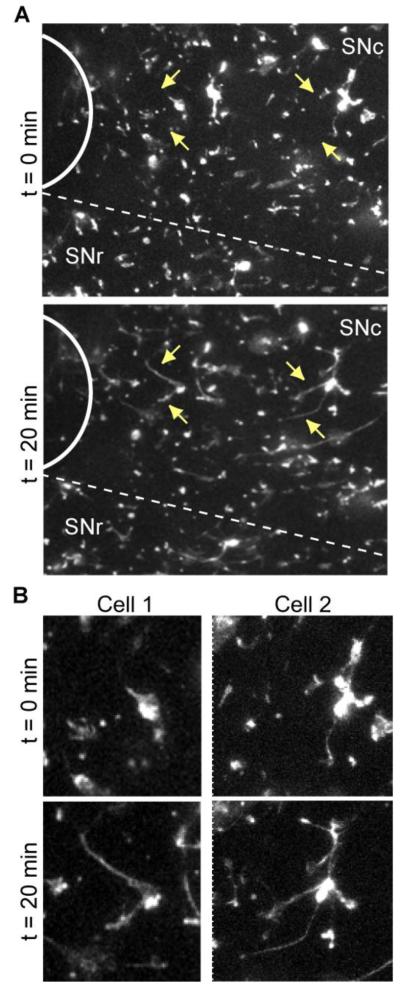
Figure 2. Response of microglia to tissue injury in acute brain slices.
Acute brain slices from CX3CR1GFP/+ were imaged with confocal microscopy over time before and after induction of tissue injury in the same slice. For image analysis, the optical stacks at each time point were converted to 2D maximum intensity projections. A, A portion from a representative slice showing microglial response to the damage immediately after (t = 0 min) and 20 min after the injury. Location of the injury is indicated with a solid white arc. The approximate border to the SNc and SNr is represented with a dashed line. Arrows point to microglial cells with processes that moved in the direction of the injury. The same cells are enlarged in B.

