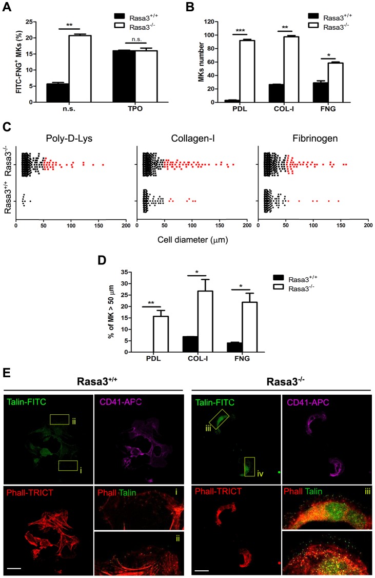
Figure 5. Altered inside-out and outside-in integrin signaling in Rasa3−/− megakaryocytes.
Fetal liver cell (FLC) were isolated from E12.5 Rasa3+/+ and Rasa3−/− embryos and cultured with TPO for 3 days. Megakaryocytes were enriched on a BSA-gradient, deprived of serum for 4 hours and used in inside-out and outside-in integrin signaling assays. A. Inside-out αIIbβ3 integrin signaling was investigated in megakaryocytes by quantifying soluble FITC-fibrinogen (FITC-FNG) bound to the CD41+ cell surface by flow cytometry. Megakaryocytes were treated with or without 100 ng/ml TPO for 30 min. Specific binding was obtained after subtraction of the amount of soluble fibrinogen bound to the cell surface in the presence of EDTA and was expressed relative to the maximum binding obtained in the presence of MnCl2. A. U.: arbitrary units. n. s.: non stimulated. B., C., D. and E. Megakaryocytes were incubated for 18 hours on Poly-D-Lysine- (PDL), collagen-I- (COL-I) and fibrinogen- (FNG) coated plates in medium containing 10% FBS. Number (B, mean ± SEM) and diameter (C) of adherent megakaryocytes (MKs) was determined in 16 fields. Results are representative of 3 independent experiments. D. The percentage of Rasa3−/− adherent megakaryocytes with a diameter over 50 µm was significantly increased, as compared with Rasa3+/+ megakaryocytes (mean ± SEM of 3 independent experiments). E. Rasa3+/+ and Rasa3−/− PDL-adherent megakaryocytes were stained with phalloidin-TRICT (actin, red), CD41-APC (magenta) and Talin-FITC (green). Confocal images were obtained from the bottom of the cells. (i–iv): 4× Digital magnification of phalloidin-TRICT and Talin-FITC merge. An increased Talin staining is observed in Rasa3−/− megakaryocytes (iii and iv), as compared with Rasa3+/+ megakaryocytes (i and ii). Scale bar: 50 µm. Statistics (unpaired t test): *: P<0.05; **: P<0.01; ***: P<0.001.

