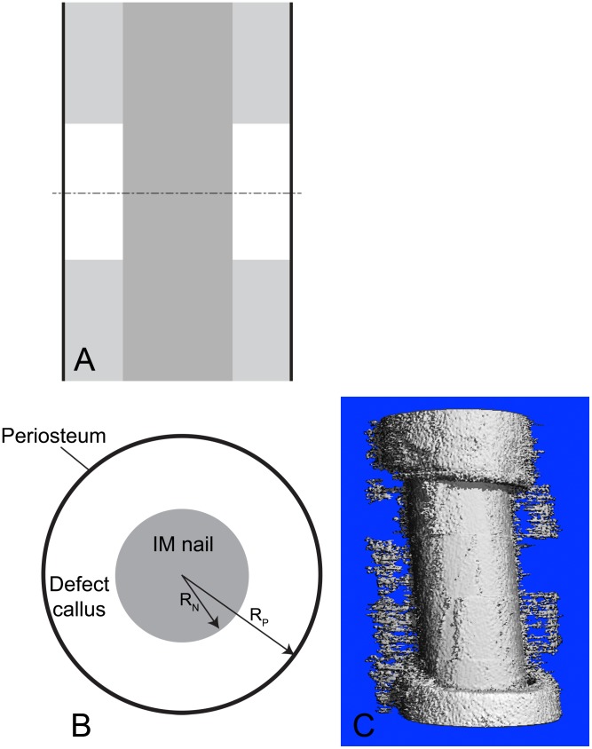
Figure 1. Prediction of bone tissue regeneration in a virtual model of a critical sized femoral defect tested in an ovine experimental model.
(A) Schematic depiction of experimental model in longitudinal plane. Proximal (upper) and distal (lower) bone are represented in light gray, with the intramedullary nail in dark gray and the periosteum (lines) in black). The middle of the defect is indicated by the cut (dashed) line, giving the transverse orthogonal plane for the radial perspective (in B). (B) Spatial system diagram for the current model. System diagram depicting nascent tissue genesis (‘callus’) in the defect, defined as the region between the intramedullary (IM) nail and the surrounding periosteum. (C) Tissue genesis in the defect proceeds predominantly from the outside in, radially from the periosteum, rather than from the proximal and proximal and distal edges (longitudinally) toward the center of the defect, as evidenced by high resolution micro-CT of actual healing in the experimental ovine defect described by the predictive model [1], [4].

