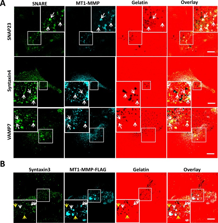
FIGURE 1:
Localization of SNAP23, Syntaxin4, VAMP7, and MT1-MMP at invadopodia. (A) Cells were plated on 594-labeled gelatin, fixed, permeabilized, and stained for SNAP23, Syntaxin4, or VAMP7 (green) along with MT1-MMP (blue). (B) Cells transfected with MT1-MMP-FLAG were plated on 594-labeled gelatin, fixed, permeabilized, and stained for Syntaxin3 (green) and FLAG epitope (for MT1-MMP, blue). Invadopodia are marked by the colocalization of MT1-MMP and dark spots of degraded gelatin. Scale bar, 10 μm.

