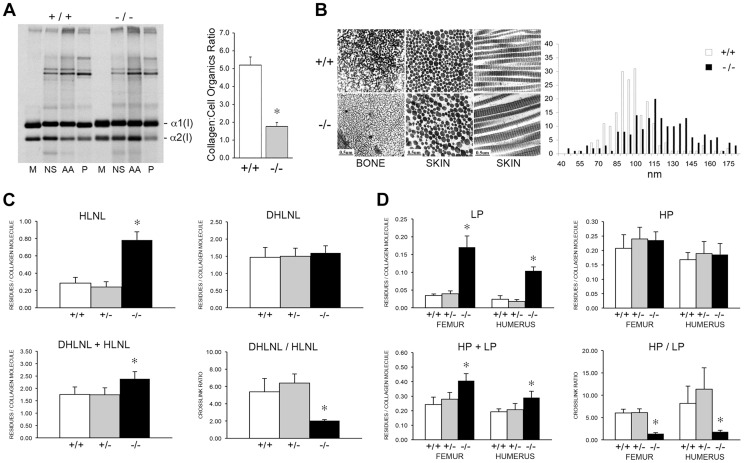
Figure 10. Dysregulation of collagen deposition and fibril assembly.
(A) Left, Deposition of type I collagen by osteoblasts into extracellular matrix in culture. Post-confluent cultures were pulsed for 24 hr, followed by serial extraction of incorporated collagens from the media (M), neutral salt (NS), acid soluble (AA, immaturely crosslinked) and pepsin soluble (P, maturely crosslinked) fractions of the matrix. Right, Matrix collagen to cell organics ratio from Raman micro-spectroscopy shows decreased collagen content in matrix deposited by homozygous Ppib-null (−/−) versus wild-type (+/+) osteoblasts in culture (p = 0.002). (B) Transmission electron micrographs of femoral and dermal collagen fibrils from 8 week-old wild-type (+/+) and CyPB-deficient mice (−/−). Diameters of 200 dermal fibrils were measured for each sample and plotted, right. (C) Quantitation of divalent crosslinks in murine humeri reveals increased HLNL (hydroxylysinonorleucine) crosslinks, which require helical lysine residues, but no change in DHLNL (dihydroxylysinonorleucine) crosslinks, which involve helical hydroxylysine residues. (D) Quantitation of trivalent crosslinks in murine humeri and femora. Total pyridinoline crosslinks are increased due to an increase in lysyl pyridinoline (LP), but not hydroxylysyl pyridinoline (HP) crosslinks in bone.

