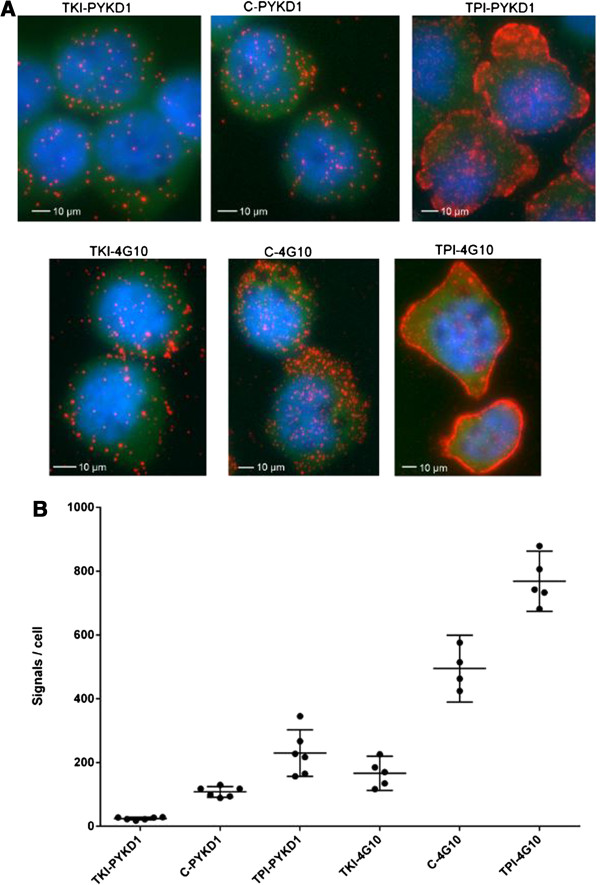
Figure 3.

In situ PLA for visualization and quantification of total pTyr levels. K562 cells probed in in situ PLA with anti-pTyr antibodies PYKD1 and 4G10. Untreated control cells, C-PYKD1 and C-4G10, are compared to cells treated with tyrosine kinase inhibitor (TKI) and tyrosine phosphatase inhibitors (TPI), respectively, to investigate whether the assay is able to measure introduced perturbations. A) Visualization by fluorescent images: Red dots represent detected pTyr signals, cell nuclei are stained in blue and cytoplasms in green. Scale bars 10 μm. B) Quantification: The number of total pTyr signals per cell in a diagram. Bars represent average with 95% confidence interval.
