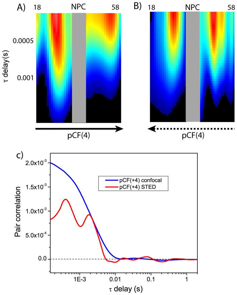
Figure 7. Confocal-pCF analysis of NLS-GFP in cells.

(A,B) Confocal-pCF(4) carpets obtained along the N→C (A) and C→N (B) directions along a line crossing the N/C interface. The two carpets are the confocal-pCF analogs of the STED-pCF carpets reported as Figure 3C,D in main text. (C) Confocal (blue) and STED-pCF(4) (red) curves for pixel 28 in the scanned line across the nuclear envelope.
