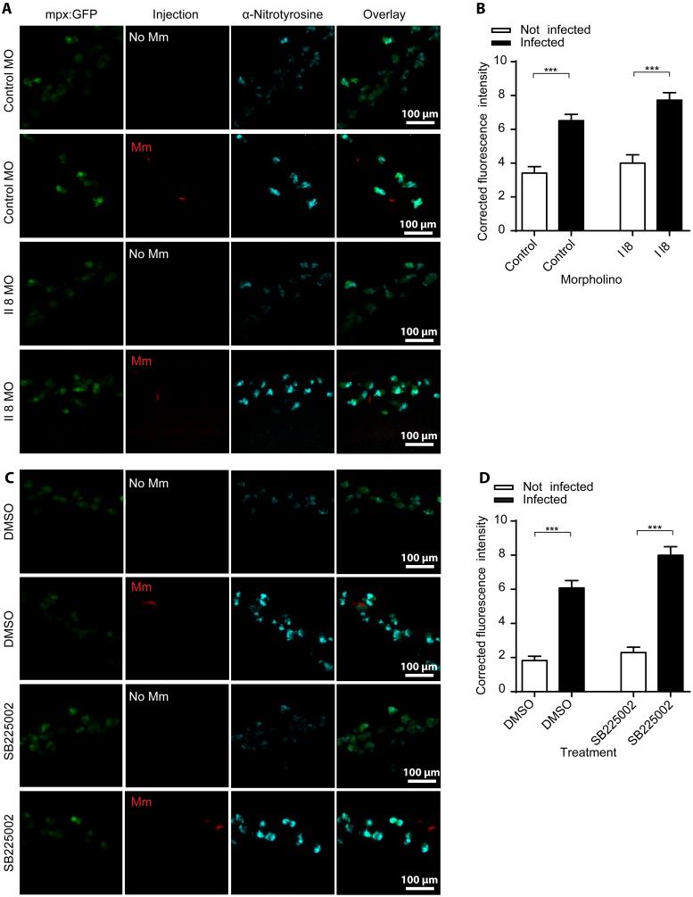
Figure 4. Increased nitrotyrosine levels in neutrophils post-infection is independent of Il8/Cxcr2 signaling.
(A) Example fluorescent confocal micrographs of the caudal vein region stained with anti-nitrotyrosine at 1 dpi, following injection of either the standard control morpholino or the il8 splice blocking morpholino at the 1-cell stage. Larvae shown are in the presence or absence of Mm, as indicated in the panels. (B) Corrected fluorescence intensity levels of anti-nitrotyrosine antibody confocal z-stacks of equal size 1 day after injection of control or il8 morpholino. Data shown are mean ± SEM, n = 90 cells from 15 embryos combined from 3 independent experiments. (C) Example fluorescent confocal micrographs of the caudal vein region stained with anti-nitrotyrosine at 1 dpi, following treatment with the Cxcr2 inhibitor SB225002 or DMSO control. Larvae shown are in the presence or absence of Mm, as indicated in the panels. (D) Corrected fluorescence intensity levels of anti-nitrotyrosine antibody confocal z-stacks of equal size 1 day after treatment of DMSO or SB225002. Data shown are mean ± SEM, n = 90 cells from 15 embryos combined from 3 independent experiments.

