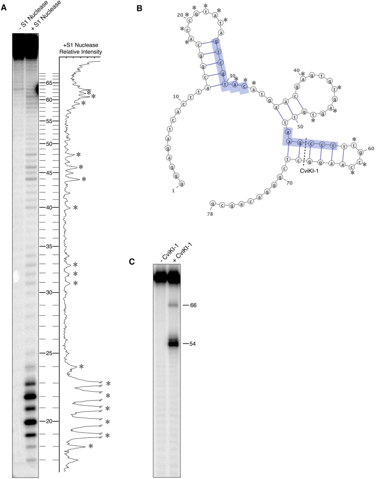
Figure 2. Secondary structure probing of aptamer-19.
A) 32P-labeled aptamer was digested with S1 nuclease and resolved by denaturing gel electrophoresis. The relative band intensities of the +S1 nuclease lane are plotted according to nucleotide position at the right of the gel. B) The secondary structure of aptamer-19 as predicted by mFold given the single stranded constraints determined by S1 nuclease digestion. Conserved motifs are highlighted in blue. C) Restriction digest with CviKI-1 confirms the presence of the third stem-loop containing the double stranded recognition sequence 5′-AGCC-3′. The CviKI-1 site is labeled in panel B.

