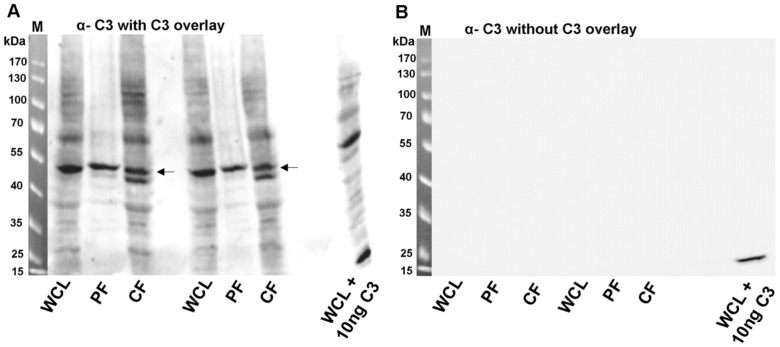
Figure 2. C3-overlay (binding of C3 to HT22 proteins).
A) Whole cell lysate, cytosolic fraction or particulate fraction from HT22 cells were generated as described in material and methods followed by separation through SDS-PAGE and transfer onto nitrocellulose. Nitrocellulose was incubated with 10 µg/ml of C3 for 60 min at 4°C. After washing bound C3 was detected by anti-C3. Arrows indicate the protein of interest (55 kDa). B) The right panel shows the anti-C3 Western blot without C3-overlay. M = molecular mass marker, WCL = whole cell lysate, PF = particulate fraction, CF = cytosolic fraction, WCL +10 ng C3 = C3 was added to whole cells lysate prior to SDS-PAGE and blotting to generate a positive C3 signal.

