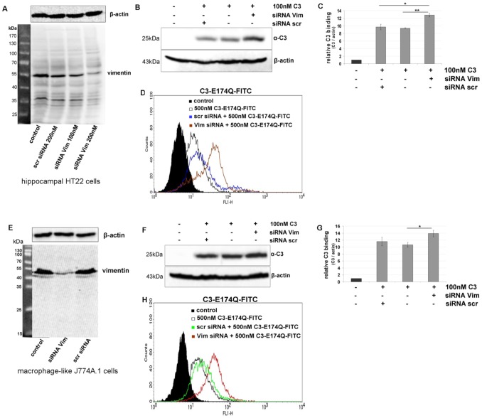
Figure 6. Knock down of vimentin in hippocampal HT22 cells and J774A.1 macrophages.
A) HT22 cells were transfected with siRNA for 48 h (scr = scrambled, Vim = vimentin). Vimentin and β-actin were detected by Western blot analysis of cell lysates. B) After siRNA transfection for 48 h, HT22 cells were exposed to C3 (100 nM) for 1 h at 4°C. Bound C3 was detected in Western blot with anti-C3. β-actin was used as internal control. C) Densitometric evaluation of bound C3 (from B) and adjustment to the corresponding actin band are shown; the bars give the relative C3 binding. D) HT22 cells transfected with siRNA for 48 h were incubated with C3-E174Q-FITC (500 nM) for 1 h at 4°C and bound C3-E174Q-FITC was analyzed by FACS cytometry. E – G) Same experiments for J774A.1 macrophages. E) Knock down of vimentin. F) Binding of C3 to cells with vimentin knock down. G) Densitometric evaluation of F. H) Binding of C3-E174Q-FITC to cells with vimentin knock down and FACS analysis.

