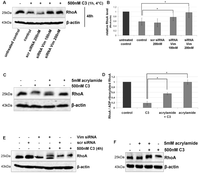
Figure 8. Uptake of C3 in HT22 and J744A.1 cells is dependent on vimentin distribution and integrity.
A) Influence of Vim-siRNA knock down (for 48 h) on the uptake of C3 into HT22 cells detected as RhoA degradation (induced by C3-catalysed ADP-ribosylation). In a pulse-chase experiment, HT22 cells were incubated with C3 (500 nM) at 4°C for 60 min. Afterwards unbound C3 was removed by washing the cells three times with PBS and fresh medium was added. Cells were then cultivated for further 48 h. Cell lysates were generated and separated by SDS-PAGE followed by Western blot analysis probing RhoA and β-actin. One representative experiment is shown (n = 3 independent experiments). B) Cellular levels of RhoA proteins were quantified by densitometric evaluation of RhoA (from A) and adjusted to the corresponding actin band. C) HT22 cells were pre-treated with acrylamide (5 mM) for 30 min followed by incubation with C3 (500 nM) for 24 h. Cells were lysed and submitted to Western blot analysis probing RhoA and β-actin. C3 alone causes a complete mol weight shift of RhoA in SDS-PAGE. Western blot analysis of one representative experiment is shown (n = 3 independent experiments). D) RhoA shift (indicative of Rho-ADP-ribosylation) by quantified by densitometric evaluation of RhoA (from C) and adjusted to the corresponding β-actin signal. E) Influence of Vim-siRNA knock down (for 48 h) on the uptake of C3 into J774A.1 cells detected as incomplete RhoA ADP-ribosylation. J774A.1 macrophages were incubated with C3 (500 nM) at 37°C for 4 h. Cell lysates were generated and separated by SDS-PAGE followed by Western blot analysis probing RhoA and β-actin. One representative experiment is shown (n = 3 independent experiments). F) J774A.1 cells were pre-treated with acrylamide (5 mM) for 30 min followed by incubation with C3 (500 nM) for 4 h. Cells were lysed and submitted to Western blot analysis probing RhoA and β-actin.

