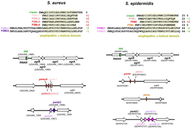
Fig. 3. PSMs in S. aureus and S. epidermidis.
Amino acid sequences are shown at the top. Numbers at the right show the net charge of the peptides at pH 7.0, rounded to whole numbers, and considering N-formylation. Genes are shown at the bottom. Gene numbering is according to strains S. aureus USA300 FPR3757 (Diep et al., 2006) and S. epidermidis RP62A (Gill et al., 2005), respectively.

