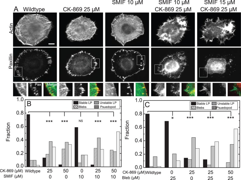
Figure 7. Myosin or Formin Inhibition does not abrogate pseudopodial protrusions in Arp2/3 inhibited cells.
(A) Images of F-actin visualized by fluorescent phalloidin (top row) and paxillin immunofluorescence (middle row) and enlargements of regions of interest (bottom row) indicated by square for wildtype MCF10A cells or cells treated with CK-869 and the formin inhibitor SMIFH2 at the indicated concentrations. (B) Fraction of MCF10A cells treated with indicated concentrations of CK-869 and SMIFH2 displaying the protrusion phenotypes as in Figure 6 (n = 41, 37, 30, 17, 65 and 44 cells respectively). Cells were treated for four hours prior to imaging. (C) Fraction of MCF10A cells treated with indicated concentrations of CK-869 and myosin ATPase inhibitor blebbistatin displaying the protrusion phenotypes as in Figure 6 (n = 55, 39, 49, 75, 26 cells respectively). Cells were treated with CK-869 for four hours prior to imaging and were treated with blebbistatin at the start of imaging. NS, not significant; *, P<0.05; ***, P<0.001 with respect to WT or control.

