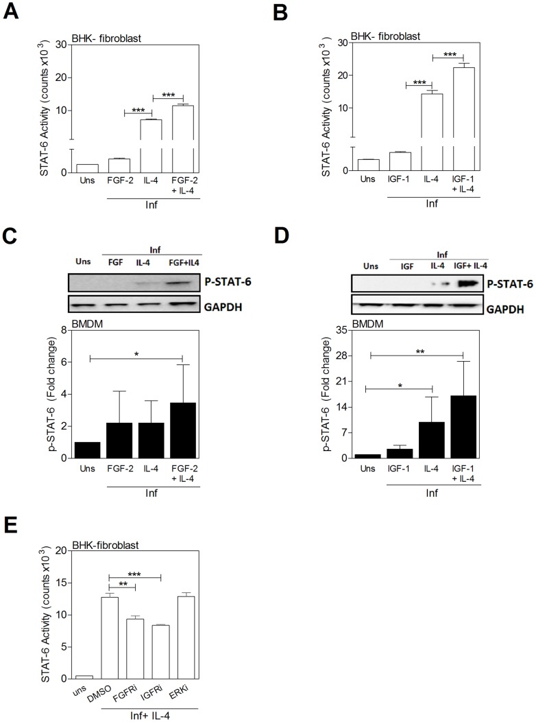
Figure 9. IL-4 and growth factors amplify STAT6 activation.
A, B) STAT6 activation measured in a luciferase reporter assay in BHK cells stimulated for 24 h with growth factors and a sub-maximal concentration of IL-4. A) IL-4 (3 IU/mL) and/or FGF-2 (20 ng/mL) and B) IL-4 (6 IU/mL) and/or IGF-1 (100 ng/mL). Shown is the mean and SEM of STAT6 activity determined by luminometry from a single experiment that was representative of 3 independent experiments. C, D) Immunoblots of cell lysates (not immunoprecipitation as shown in Fig. 8B) showing phospho-STAT6 in BMDMs stimulated for 20 min with L. donovani promastigotes and C) IL-4 (8 IU/mL) and/or FGF-2 (20 ng/mL) or D) IL-4 (20 IU/mL) and/or IGF-1 (100 ng/mL). Bars represent the mean and SEM of fold change with reference to the uninfected, unstimulated (Uns) controls calculated by densitometry analysis of immunoblot bands from 3 independent experiments. Also shown is a representative individual immunoblot. E) IL-4-mediated activation of STAT6 in infected BHK fibroblasts is reduced by inhibition of FGFR and IGF-1R but not ERK. The cells were exposed to the control (DMSO) or FGFR inhibitor (FGFRi; PD166866; 10 µM), IGFR inhibitor (IGFRi; PPP; 5 µM), or ERK inhibitor (ERKi; PD98059; 5 µM) for 1 hr and then stimulated for another 24 hrs with IL-4 (25 IU/mL) in the presence or absence of inhibitor. Shown is the mean and SEM of STAT6 activity determined by luminometry from a single experiment that was representative of 2 independent experiments.

