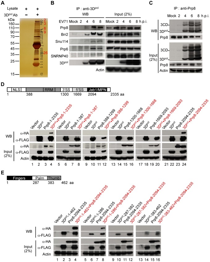
Figure 1. 3Dpol associates with the nuclear protein Prp8.
(A) Identification of potential 3Dpol-interacting host proteins. The cell lysates for IP were harvested from EV71 40 MOI-infected RD cells at 6 h.p.i. and treated with the 3Dpol monoclonal antibody or untreated as a negative control. The proteins that interacted with 3Dpol were pulled down using an anti-3Dpol antibody, along with protein A-Sepharose, and detected by 1D SDS-PAGE and silver staining. (B) 3Dpol interacts with 5 components of U5 snRNPs, including Prp8, Brr2, Snu114, Prp6, and SNRNP40. The interaction of EV71 3Dpol and the nuclear protein U5 snRNPs was further confirmed by Co-IP and WB assays. The lysates harvested from mock- or EV71 40 MOI-infected RD cells at 2 to 8 h.p.i. were treated with RNase A (10 µg/ml) and immunoprecipitated using an anti-3Dpol antibody. The 5 components of the U5 snRNPs that interacted with 3Dpol were detected using a WB assay. The input samples were verified in the presence of 3Dpol and the five components of the U5 snRNPs in the lysates. Actin served as an internal control. (C) The core spliceosome splicing factor Prp8 can also pull down 3Dpol and 3CD. EV71-infected RD cell lysates from 2 to 8 h.p.i. were treated with RNase A (10 µg/ml) and incubated with antibodies against the Prp8 probe. After the IP assay, 3Dpol and 3CD were analyzed using WB with an anti-3Dpol antibody. (D) 3Dpol associates with the C-terminal domain of Prp8 containing the Jab1/MPN region. The functional domain architecture of human Prp8 is shown (upper panel). HEK293T cells were transfected with plasmids encoding full-length FLAG-3Dpol (lanes 2, 4, 6, 8, 10, 12, 14, 16, 18, 20, 22, and 24), various truncated forms of HA-Prp8 (lanes 3, 4, 7, 8, 11, 12, 15, 16, 19, 20, 23, and 24), and empty vectors (lanes 1, 5, 9, 13, 17, and 21). At 48 h after transfection, the lysates were treated with RNase A (10 µg/ml) and immunoprecipitated with antibodies against FLAG. The truncated form of Prp8 that interacted with 3Dpol was detected by WB using an antibody against HA. (E) The C-terminal domain of Prp8 interacts with the fingers domain of 3Dpol. The functional domain architecture of EV71 3Dpol is shown (upper panel). HEK293T cells were transfected with plasmids encoding HA-Prp8-2094-2335 (lanes 3, 4, 7, 8, 11, 12, 15, and 16), various truncated forms of FLAG-3Dpol (lanes 2, 4, 6, 8, 10, 12, 14, and 16), and empty vectors (lanes 1, 5, 9, and 13). The various truncated forms of 3Dpol were pulled down by IP with an anti-FLAG antibody. The C-terminal domain containing the Jab1/MPN region of Prp8, which interacts with the truncated form of FLAG-3Dpol, was detected with an anti-HA antibody in a WB assay.

