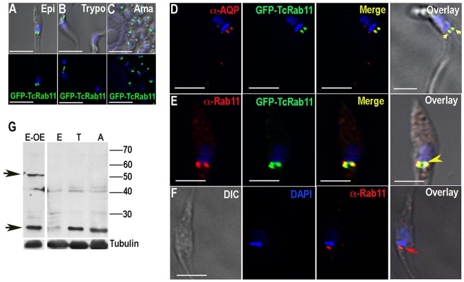
Figure 1. Fluorescence microscopy analysis of TcRab11 in different stages of T. cruzi.
(A–C) GFP fusion protein of TcRab11 was detected in the contractile vacuole bladder of epimastigotes (Epi, A), trypomastigotes (Trypo, B), and intracellular amastigotes (Ama, C) using antibodies against GFP. Upper panels show differential interference contrast microscopy (DIC) images merged with DAPI staining of DNA (in blue) and GFP-TcRab11 (in green). Lower panels show fluorescence images. (D) GFP-TcRab11 (green) co-localizes with antibodies against T. cruzi aquaporin 1 (α-AQP, red), a marker for the contractile vacuole, under hyposmotic conditions. (E) Antibodies against TbRab11 (α-Rab11, red) co-localize with GFP-TcRab11 (green). (F) Antibodies against TbRab11 (red) localize to a compartment that resembles the contractile vacuole in (E). DAPI staining is in blue. Arrowheads in D–F show co-localization between antibodies against TcAQP1 and GFP (D), TbRab11 antibody and GFP (E) and labeling with antibodies against TbRab11 (F), respectively. Bars in A–F = 10 µm. (G) Western blot analyses with TbRab11 antibody of lysates of epimastigotes overexpressing GFP-TcRab11 (E-OE), or wild-type epimastigotes (E), trypomastigotes (T) and amastigotes (A) showing bands (arrows) corresponding to the endogenous TcRab11 (24 kDa) and to GFP-TcRab11 (50 kDa). The blots were sequentially probed with αTbRab11 and anti-tubulin antibodies, used as loading control.

