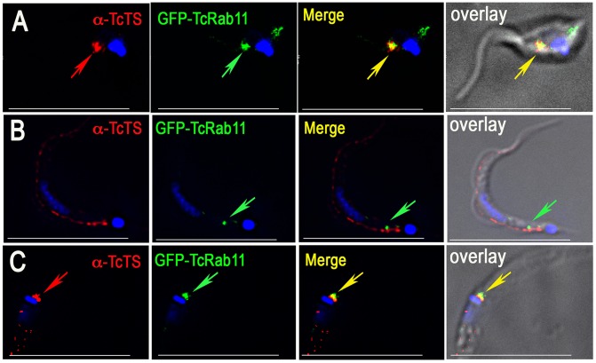
Figure 5. Localization of TcTS during differentiation to cell-derived and metacyclic trypomastigotes.
(A) Co-localization of αTcTS and antibodies against GFP in intermediate stages (epimastigote-like) obtained from tissue culture supernatants. (B) TcTS localizes to patches of the plasma membrane in fully differentiated trypomastigotes while GFP-TcRab11 remains in the CVC. (C) Co-localization of αTcTS with GFP-TcRab11 in epimastigotes during transformation into metacyclic stages. Scale bars (C–E) = 10 µm.

