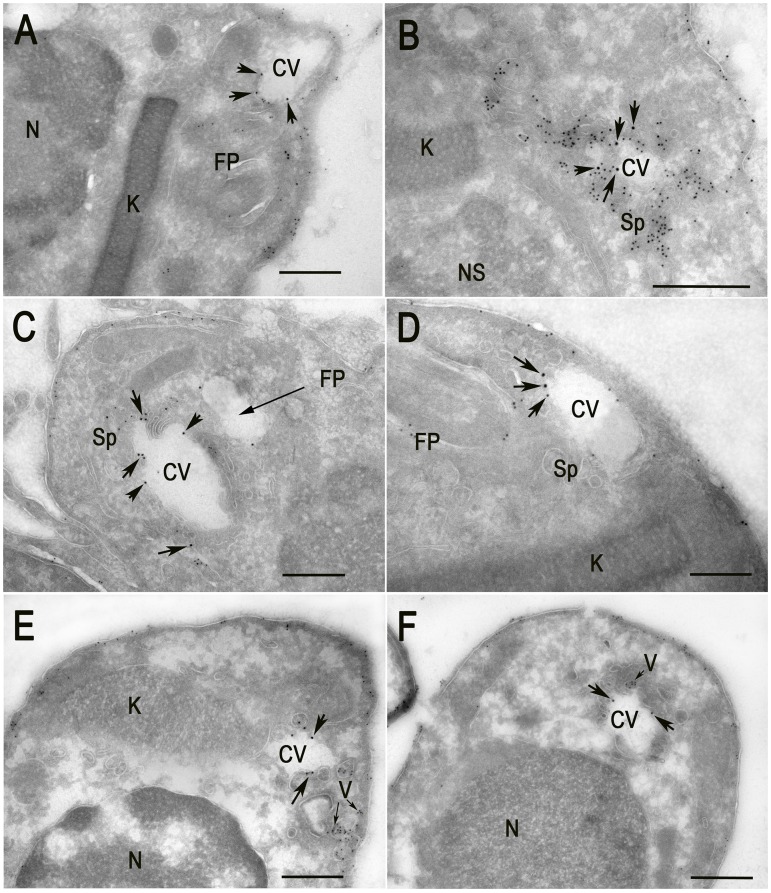
Figure 6. Cryo-immunoelectron microscopy localization of GFP-TcRab11 and TcTS in amastigotes.
Amastigotes were isolated from HFF at different times p.i., as described under Materials and Methods. GFP-TcRab11 and TcTS were detected with goat anti-GFP, and rabbit anti-TcTS antibodies, and donkey anti-goat 18 nm colloidal gold and donkey anti-rabbit 12 nm colloidal gold, respectively. (A–D) Amastigotes obtained after 96 h p.i. Co-localization of antibodies against GFP (arrows) and TcTS (small dots) is evident in the CV bladder (CV) and spongiome (Sp), while TcTS also localizes to the flagellar pocket (FP) and in patches of the plasma membrane. Note in (B) a collapsed bladder and intense labeling of the spongiome. (E–F) Amastigotes obtained 106 h p.i. GFP-TcRab11 localizes to the CV bladder while TcTS localizes to vesicles (V, small arrows) close to the plasma membrane and in patches in the plasma membrane. Scale bars = 500 nm. Note that the patchy appearance of the cytoplasm is due to the absence of glutaraldehyde in the fixative because it abolished labeling of TcTS.

