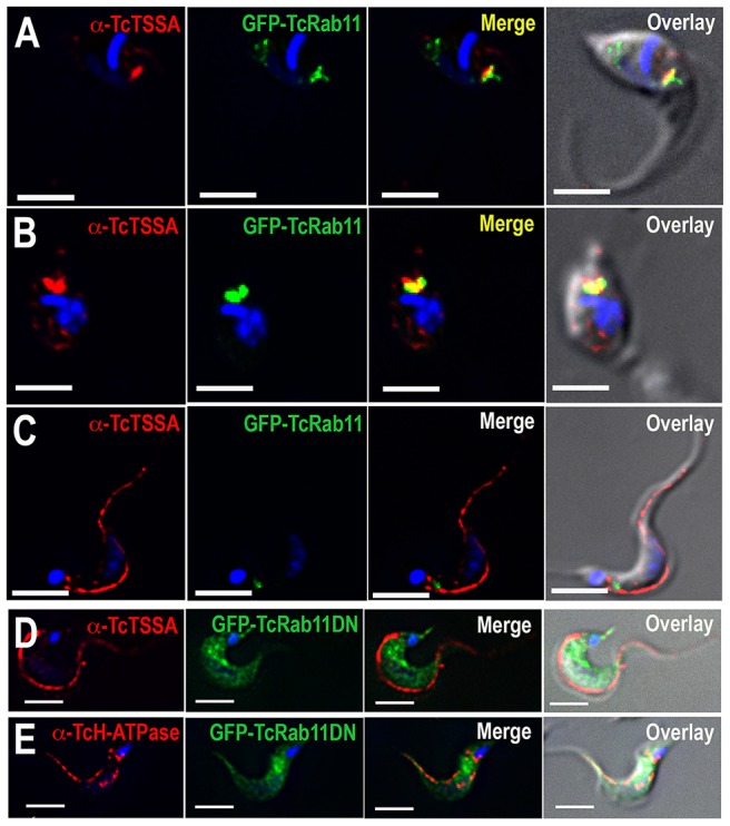
Figure 8. Localization of surface proteins in GFP-TcRab11OE and GFP-TcRab11DN-expressing parasites.

Antibodies against TcTSSA II (red) co-localize with antibodies against GFP (green) in intermediate forms (A) and amastigotes (B) but not in trypomastigotes expressing GFP-TcRab11, where they localize to the plasma membrane (C). Antibodies against TcTSSA II (D) still localize to the plasma membrane in GFP-TcRab11DN-expressing cells, while antibodies against the H+-ATPase (E) maintain their intracellular and plasma membrane localization in GFP-Rab11DN-expressing cells. In (D) and (E) GFP staining localizes to the cytosol. Scale bars = 10 µm.
