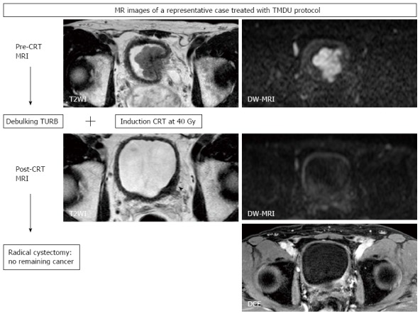
Figure 6.

Magnetic resonance images of a 61-year-old man with muscle-invasive bladder cancer (urothelial cancer, stage cT3, grade 3) treated with the Tokyo Medical and Dental University protocol consisting of transurethral resection of bladder tumor and induction chemoradiotherapy (CRT) followed by radical or partial cystectomy. T2WI shows a large hypointense tumor at the bladder neck, invading the prostate. At the diagnosis, DW-MRI with a b-value of 1000 s/mm2 displays a hyperintense lobulated mass. After TURBT and CRT, this lesion shows wall thickening (arrow head) on T2WI and enhancement on DCE, while the abnormal signal on DW-MRI is diminished to normal signal intensity. Histopathologic examination of the cystectomized sample reveals no remaining bladder cancer, revealing the findings of post-CRT T2WI and DCE to be false-positive findings reflecting post-treatment changes in bladder tissues. TURBT: Transurethral resection of bladder tumor; CRT: Chemoradiotherapy; DW-MRI: Diffusion-weighted magnetic resonance imaging; DCE: Dynamic contrast-enhanced.
