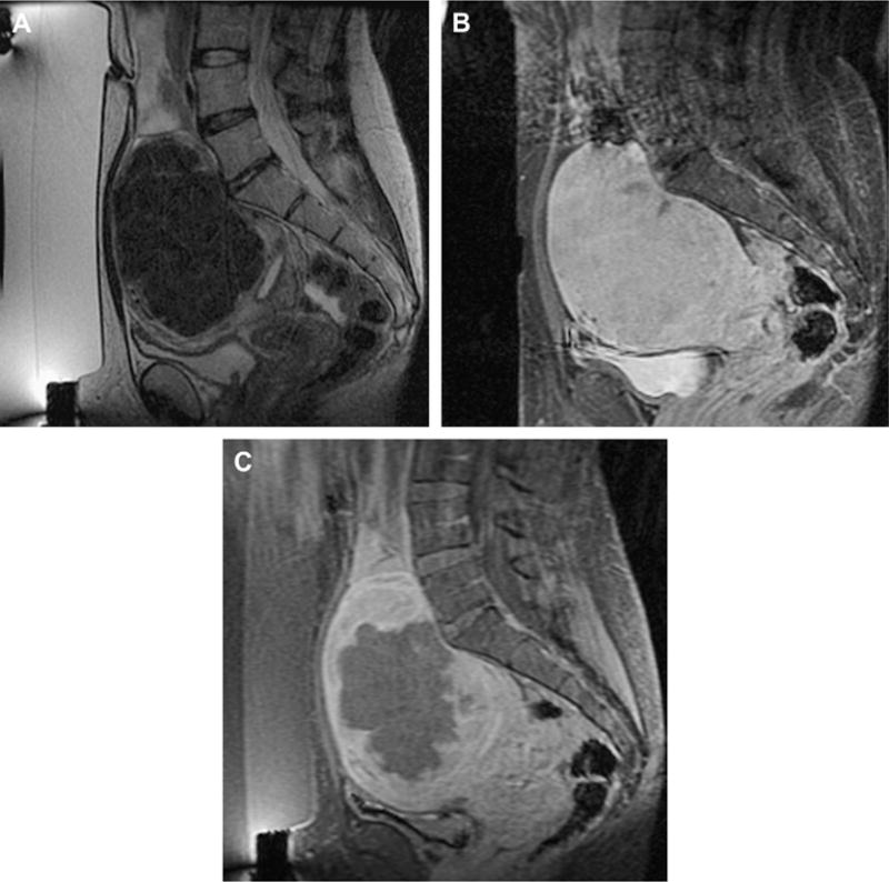
Fig. 3.

Imaging of a uterine fibroid pretreatment (A, B) and post-treatment (C) with MRgFUS. Sagittal T2-weighted image (A), obtained with the patient in the prone position overlying the US transducer, demonstrates a large solitary uterine fibroid of low-signal intensity. Sagittal SPGR post gadolinium (B) demonstrates homogenous enhancement of the fibroid. After treatment, sagittal SPGR post gadolinium (C) demonstrates a new large nonperfused area within the fibroid, consistent with treatment-induced necrosis.
