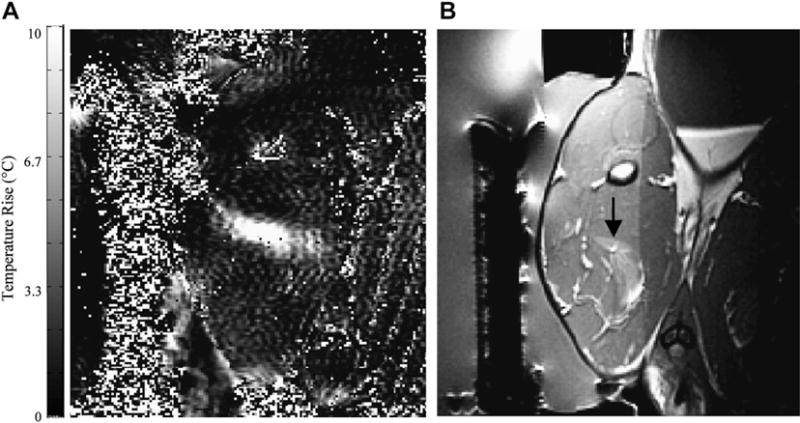
Fig. 8.

(A) MR imaging–based temperature image during a sonication (130 W for 30 seconds) into rabbit thigh muscle during a test of an MR imaging–compatible transrectal phased array applicator for MRgFUS of prostate. (B) The thermal lesion (arrow) seen in T2-weighted imaging. The bright region to the right of the lesion is a tissue fascia layer. (From Sokka SD, Hynynen K. The feasibility of MRI-guided whole prostate ablation with a linear aperiodic intracavitary ultrasound phased array. Phys Med Biol 2000;45:3373–83; with permission.)
