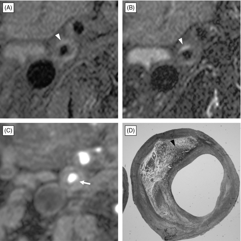
Fig. 1.
Atherosclerotic plaque composed of fibrous tissue and small amount of intraplaque hemorrhage with no fibrous cap rupture. (A) black-blood fat-suppressed T1-weighted image, (B) black-blood fat-suppressed T2-weighted image, (C) source image of time-of-flight magnetic resonance (TOF MR) angiogram, (D) histological section with Masson-Trichrome staining. Black-blood fat-suppressed T1-weighted image (A) and T2-weighted image (B) demonstrates carotid plaque composed of a large isointensity portion and small crescent-shape high intensity (arrowhead) relative to the submandibular gland. Source image of TOF MR angiography (C) shows moderate stenosis of the right carotid artery with thin hypointensity band of fibrous cap covering the plaque completely. Note partial thinning (arrow) on the fibrous plaque. Histological section with Masson-Trichrome staining (D) demonstrates fibrous cap covering the fibrous plaque with intraplaque hemorrhage (arrowhead), and no disruption is found.

