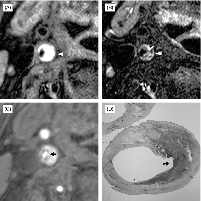
Fig. 3.
Ulcerated atherosclerotic plaque composed of lipid core and intraplaque hemorrhage with rupture of fibrous cap. (A) black-blood fat-suppressed T1-weighted image, (B) black-blood fat-suppressed T2-weighted image, (C) source image of time-of-flight magnetic resonance (TOF MR) angiogram, (D) histological section with Masson-Trichrome staining. The atherosclerotic carotid plaque demonstrates a large portion of high signal intensity on black-blood fat-suppressed T1-weighted image (A) and mixed high and intermediate signal intensity on black-blood fat-suppressed T2-weighted image (B). Source image of TOF MRA (C) shows disruption of thick fibrous cap (arrow) covering the hyperintense carotid plaque. Histopathological examination with Masson-Trichrome staining (D) clearly reveals that a plaque composed of lipid core and intraplaque hemorrhage has small ulceration with disruption of fibrous cap (arrow). Note protruded lumen (arrow) into hyperintense plaque on black-blood fat-suppressed T2-weighted image (B).

